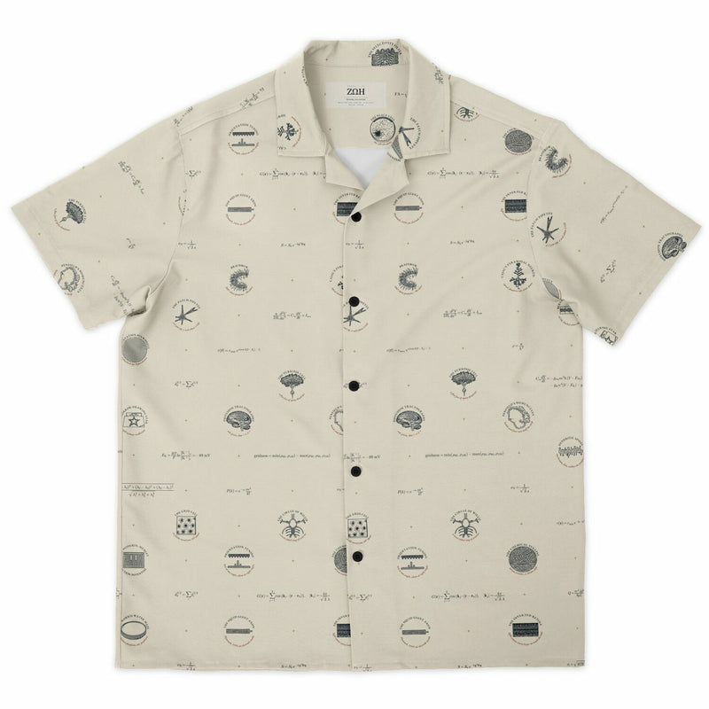
NEURON DOCTRINE
Cajal’s Pyramidal Neuron
apical dendrite up, axon down
Golgi’s stain blackens perhaps one neuron in a hundred, but completely, so Cajal could draw single cells whole. He called the cortical pyramidal cell the psychic cell: a triangular body, one apical dendrite climbing to a tuft, a skirt of basal dendrites and one axon descending to the white matter. Only the dendrites carry spines.
NEURON DOCTRINE
The Chemical Synapse
40 nm vesicles, a 20 nm cleft
At the frog neuromuscular junction in 1954, del Castillo and Katz found transmitter released in packets of fixed size. With calcium lowered, many stimuli released none, and the counts fitted Poisson’s distribution, so the failures alone gave the mean. In 1979 Heuser, Reese and colleagues froze synapses within milliseconds of a stimulus and caught the vesicles opening.
NEURON DOCTRINE
The Purkinje Cell
a flat fan of spiny dendrites
Jan Purkyně described these flask-shaped cells in 1837. The dendritic tree is flat, a few micrometers thick and hundreds wide, threaded at right angles by parallel fibers whose synapses are many and weak. One climbing fiber wraps the main dendrites and fires a complex spike almost every time. The axon is the cerebellar cortex’s only output, and it inhibits.
NEURON DOCTRINE
Dendritic Spines
mushroom, thin, stubby: one synapse each
Cajal saw these thorns in 1888 and insisted they were real; in 1959 E. G. Gray found synapses on them. Almost every spine carries one excitatory synapse, and head volume scales with synapse area, so shape reads out strength. A neck 1 µm long and 0.1 µm wide, with a resistance of about 127 MΩ, makes each spine a compartment.
NEURON DOCTRINE
The Node of Ranvier
sodium channels packed into 1 µm
Ranvier described the gaps in 1878, and Tasaki showed in 1939 that the impulse leaps between them. Sodium channels crowd the node at over a thousand per square micrometer. In 1951 Rushton worked out that for a fiber of fixed width, conduction is fastest when the axon is 1/√e, about 0.607, of the fiber’s diameter.
ALL OR NONE
The Patch Pipette
a 1 µm tip, a seal above 1 GΩ
In 1976 Neher and Sakmann first recorded the current through a single channel: steps of a few picoamperes, nearly swamped by the noise of a 50 MΩ seal. By 1980 gentle suction fused glass to membrane above a gigohm, and a seal’s noise falls as one over the square root of its resistance. They shared the 1991 Nobel Prize.
ALL OR NONE
The Selectivity Filter
K⁺ in single file, water between
At body temperature, 140 mM potassium inside a neuron and 5 mM outside give a Nernst potential of −89 mV. Roderick MacKinnon’s 1998 structure of KcsA showed how the channel prefers potassium to smaller sodium a thousand to one: four rings of carbonyl oxygens mimic potassium’s shell of water, and sodium does not fit. He shared the 2003 Nobel Prize.
ALL OR NONE
Channelrhodopsin-2
blue light at 470 nm opens it
Channelrhodopsin-2 comes from the eyespot of the green alga Chlamydomonas reinhardtii, which steers by it. Blue light near 470 nm opens it within a millisecond. In the brain the light arrives down a fiber 200 µm across whose numerical aperture, 0.39, sets the cone: 16.7 degrees each side in tissue, before scattering blurs it.
ALL OR NONE
The Hodgkin–Huxley Action Potential
sodium first, potassium after
Integrated here with Hodgkin and Huxley’s own parameters along a cable of their fiber at 18.5 °C, the impulse travels at 18.70 m/s, against the 18.8 Huxley reached by hand and the 21.2 they measured. Sodium conductance peaks with the spike; potassium peaks 0.39 ms later, outlasts it and makes the undershoot.
ALL OR NONE
The Squid Giant Axon
Plymouth, 1939: an electrode inside
J. Z. Young identified the giant axons in 1936, fibers up to a millimeter across that drive the squid’s escape jet. In 1939 Hodgkin and Huxley threaded a capillary down one and saw the inside swing some 40 mV positive during the impulse, overturning Bernstein’s theory that the potential should merely fall to zero.
YOU ARE HERE
The Grid Cell
six fields around one, 60° apart
In 2005 the Mosers’ group found cells in the medial entorhinal cortex that fire on a triangular lattice covering the whole floor. Three plane waves at 60 degrees reproduce the pattern: this cell’s fields sit 40 cm apart, and its gridness score is 1.28. Spacing grows by about 1.42 from one module to the next, close to √2.
YOU ARE HERE
Brainbow
2007: three proteins, ~90 colors
Brainbow turns Cre recombinase into a dice roll: in each cell it picks one of three fluorescent proteins, and several copies of the construct give every neuron its own mixture. With k copies there are (k + 1)(k + 2)/2 mixtures, 91 for twelve. The 2007 paper resolved about ninety colors, enough to follow hundreds of neighboring axons.
YOU ARE HERE
The Place Cell
one cell, one place
In 1971 O’Keefe and Dostrovsky found hippocampal cells that fire in one place, whichever way the rat faces. Muller and Kubie’s test arena is a gray cylinder with one white cue card covering 100 degrees of wall: rotate the card and the fields rotate with it. In sleep the same sequences replay, about twentyfold faster.
YOU ARE HERE
The Morris Water Maze
a hidden platform, one quadrant
Morris introduced the task in 1981: a round pool made opaque, a small platform just beneath the surface, and only the room to steer by. In 1986 he and colleagues showed that blocking NMDA receptors with AP5 prevents the learning, tying long-term potentiation to spatial memory. A 10 cm platform covers 0.44% of a 150 cm pool.
YOU ARE HERE
The Mirror-Drawing Star
skill without recollection
William Scoville removed most of both medial temporal lobes from Henry Molaison in 1953, and H.M. could no longer form lasting new memories of events or facts. In 1962 Brenda Milner had him trace a star seen only in a mirror. His errors fell over three days as anyone’s do, yet each day the task was new to him.
RECEPTIVE FIELD
The Trichromatic Cone Mosaic
L:M about 2:1, S about 1 in 20
Roorda and Williams imaged living cone mosaics in 1999 and found long- and middle-wavelength cones scattered at random; their two subjects’ L:M ratios were 1.15 and 3.79. Short-wavelength cones make up only 5 to 7 percent. At the foveal peak of 199,000 per square millimeter, cones sit 2.41 µm apart: a limit of about 70 cycles per degree.
RECEPTIVE FIELD
Orientation Tuning
best at one angle, silent at right angles
In 1958 a cell in a cat’s visual cortex burst into activity as Hubel and Wiesel slid a glass slide into their projector: the slide’s faint edge was the stimulus. Such cells answer a bar at one angle and fall silent at right angles. They listened to them through a loudspeaker, and shared the 1981 Nobel Prize.
RECEPTIVE FIELD
Orientation Pinwheels
π pinwheels per hypercolumn
Hubel and Wiesel predicted that preferred orientation would change smoothly across the cortex. Imaged optically, it swirls around points where every orientation meets, which Bonhoeffer and Grinvald named pinwheels in 1991. Kaschube, Wolf and colleagues counted 3.14 per hypercolumn area in three lineages of mammal whose columns evolved separately: π, within error.
RECEPTIVE FIELD
The Inverted Retina
light crosses every layer to reach the rods
Cajal mapped the retina’s layers in 1892: photoreceptors to bipolar cells to ganglion cells, with horizontal and amacrine cells working sideways. Light crosses them all first. Where ganglion-cell axons gather to leave the eye there are no receptors, the blind spot; at the fovea the inner layers are swept aside so light meets the cones directly.
RECEPTIVE FIELD
Ocular Dominance Columns
one eye per stripe, 0.4 mm
In 1974 Wiesel, Hubel and Dan Lam injected radioactive proline into one eye of a monkey; it traveled to layer 4C, and dark-field autoradiographs showed that eye’s input as silver stripes. With Simon LeVay they found them about 0.4 mm wide, meeting the V2 border at right angles. Close one eye early and the other’s stripes widen.
ENCHANTED LOOM
Phineas Gage’s Skull
in under the left cheekbone, out the top
On 13 September 1848 a blasting charge drove Phineas Gage’s tamping iron, 109 cm long and 3.2 cm thick, in under his left cheekbone, behind the left eye and out through the top of his skull. He survived, but John Harlow’s 1868 report described a man so changed that his friends said he was no longer Gage.
ENCHANTED LOOM
The Circle of Willis
a seven-sided ring of arteries
Two internal carotids and two vertebral arteries supply the brain, and the ring at its base links them through seven segments, so blood can be rerouted if one carotid narrows slowly. By Poiseuille’s law a communicating artery half the usual width carries a sixteenth of the flow. Fewer than half of brains have the complete ring.
ENCHANTED LOOM
Penfield’s Homunculus
hands and face: over half the strip
Penfield stimulated the exposed cortex of patients under local anesthetic and asked what they felt. With Boldrey in 1937 and Rasmussen in 1950 he drew the body along the strip behind the central sulcus, toes on the midline wall, throat by the lateral fissure. In this schematic, hand and fingers take 24% of the strip, face and mouth 39%.
ENCHANTED LOOM
Diffusion Tractography
red, green, blue = x, y, z
Water diffuses several times faster along axons than across them. Basser, Mattiello and Le Bihan fitted a tensor in every voxel in 1994, and following its main axis from voxel to voxel draws streamlines, colored by Pajevic and Pierpaoli’s code. They are an inference, not axons: they cannot show which way signals run, and crossing fibers fool simple methods.
ENCHANTED LOOM
Berger’s Alpha Rhythm
10 Hz, blocked when the eyes open
Hans Berger recorded the first human EEG in Jena in 1924 and published in 1929. Its most prominent rhythm, alpha, runs at 8 to 12 waves a second over the back of the head with the eyes closed, and vanishes when they open. At the standard paper speed of 30 mm/s, a 10 Hz alpha wave is 3 mm long.