Genetics & Molecular Biology · Subject 22

Genetics & Molecular Biology

Button-ups, ties and socks from Mendel’s 3:1 ratio to the lac operon.

Genetics & Molecular Biology

An X-gal plate, drawn for this page: blue colonies carry the empty vector, white ones the insert.

This week in Genetics & Molecular Biology,

BRCA1 variants that read differently by cell type, embryo nuclei that soften to wake their genes, cells that ride out polymerase shortfalls, and flies that defy genomic forecasts.

Get it by email

Download MP3

Behind this page is an agarose gel on an ultraviolet light box, and scrolling runs the current. Phage lambda DNA, 48,502 base pairs, cut with HindIII, is loaded in two lanes, with plasmid pBR322, cut once, between them. Each fragment moves toward the anode at a speed that falls with the logarithm of its length, and at each section of the page a band of the ladder comes to rest on the reading line: 125 base pairs at the introduction, then 564, 2,027, 4,361 (level with the 4,361 base pair plasmid), 6,557 and 9,416 at the five designs. In the stripe section the gel gives way to a Sanger sequencing film on a light box, read from the bottom up in four lanes, G, A, T and C: the first 27 bases of human beta-globin, ATG GTG CAT CTG ACT CCT GAG GAG AAG, whose seventh codon, GAG, becomes GTG in sickle-cell disease. After the stripes the gel finishes its run at the bottom of the page. The readout gives the length the law puts on the reading line, that band’s share of the lambda genome and how far the run has gone; on the film, the bases read and the last codon. The spacing follows the semi-log law of agarose electrophoresis, a model.

Welcome in

For anyone who has picked a white colony

You know the hum of the thermal cycler, the smell of LB, and the moment the UV box lights a gel and the band is exactly where you hoped. You have counted flies under CO₂ with a paintbrush, or peas, or plaques, and run the chi-square to see whether three to one was really three to one. You have read a sequence base by base and found the one letter that mattered. You remember the first white colony on a plate of blue, picked with a toothpick and grown overnight, and the miniprep that showed your insert at the right size.

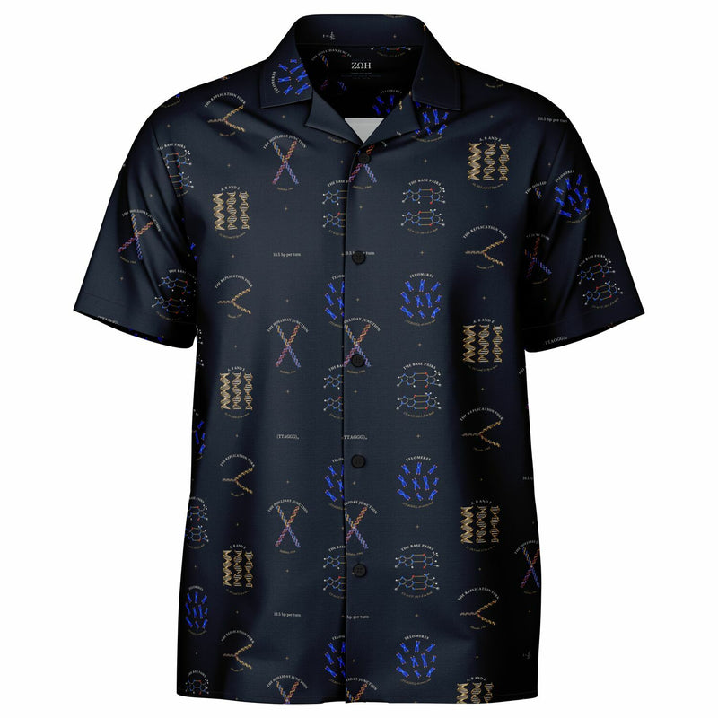
Short-sleeve button-up · XS–4XL

5′ TO 3′

How DNA pairs, copies, recombines and ends.

Adenine pairs with thymine by two hydrogen bonds, guanine with cytosine by three, and the two pairs are the same width, their sugar attachment points about 10.5 Å apart, so any sequence fits one regular helix. The strands run in opposite directions and DNA polymerase adds only to a 3′ end, so at a replication fork one new strand is made continuously and the other backward in short pieces, which Okazaki’s group found in 1968.

Holliday proposed in 1964 that recombining molecules swap strands through a four-way junction; with magnesium it folds into a stacked X. Ordinary replication cannot copy the very end of a chromosome, so vertebrate chromosomes end in long runs of TTAGGG that telomerase, found in 1985, rebuilds. And the helix comes in three classic forms: B with 10.5 base pairs a turn and A with about 11, both right-handed, and Z, left-handed, with 12.

  • The Base PairsA·T with two gold hydrogen bonds, G·C with three, and both exactly the same width: why any sequence fits the helix.
  • The Replication ForkA replication fork: one new strand continuous, the other in short gold pieces, each begun from a red RNA primer.
  • The Holliday JunctionFour arms of DNA meeting in a stacked X, two strands crossing over at the center: recombination caught midway.
  • TelomeresBlue metaphase chromosomes with a bright dot at the tip of every chromatid: the TTAGGG caps, lit by a probe.
  • A, B and ZA-, B- and Z-DNA side by side at one height: two right-handed, one left-handed, with 11, 10.5 and 12 base pairs a turn.
Shop now · Regular price $65 Sale price $58.50

Leave your email and we’ll write the day it’s ready.

Short-sleeve button-up · XS–4XL

CIS AND TRANS

How a gene is switched on, read, cut to length and packed away.

Jacob and Monod’s lac operon, in 1961, gave gene regulation its grammar: an operator acts only on the DNA it sits in, in cis, while a repressor acts on any copy, in trans. The repressor, four identical chains, grips two operators and loops the DNA between them, and lets go when allolactose binds. Spread for the electron microscope in 1969, genes being transcribed look like feathers, their RNA chains short at the start of each gene and long at the end.

Most human genes are interrupted. Splicing ties each intron into a lariat through a 2′–5′ bond at one adenosine, seen in 1984, and joins the exons. In the nucleus, DNA wraps 1.65 left-handed turns, 147 base pairs, around each core of eight histones. And about three hours after laying, a fly embryo already shows even-skipped in seven stripes, each set by its own enhancer.

  • The Lac OperonLac repressor as a glass V, its two tips gripping two operators and looping the DNA between: the switch held off.
  • The Christmas TreeThree ribosomal genes being read at once: feathers of RNA, short to long, along one DNA axis, bare spacers between.
  • The LariatThe spliced-out intron as a lasso, tied by one gold adenosine, beside the two exons it leaves joined.
  • The NucleosomeEight histones in four colors, with DNA wound 1.65 left-handed turns around them: chromatin’s repeating bead.
  • Seven StripesA fly embryo three hours old, stained for even-skipped: seven brown stripes, the first sign of the body plan.
Shop now · Regular price $65 Sale price $58.50

Leave your email and we’ll write the day it’s ready.

In the incubator: Mendel’s pods, the dihybrid tray, the Hardy–Weinberg square, the ABO tile and Johannsen’s beans.
In the incubator

THREE TO ONE

Mendel’s ratios, and what became of them in people and populations.

Mendel counted. In 1866 he reported 5,474 round seeds and 1,850 wrinkled from his hybrid peas, 2.96 to 1, and from round yellow crossed with wrinkled green, 315, 108, 101 and 32: nine, three, three and one. Each gamete carries one allele of each gene, and unlinked genes sort independently. The wrinkled allele itself was cloned in 1990: a 0.8 kb insertion, like a transposon, in the gene for starch-branching enzyme I.

In 1908 Hardy and Weinberg carried the ratios into populations: with random mating, genotypes settle at p², 2pq and q² and stay there, so dominance alone changes nothing. In 1924 Bernstein showed that the ABO groups are three alleles of one gene, A and B codominant. And Johannsen’s pure lines of beans, where selecting heavy or light seed did nothing, separated the inherited from the rest and gave the field, in 1909, the words gene, genotype and phenotype.

  • Mendel’s PeasThree dry pods from one hybrid plant: eighteen round peas and six wrinkled, side by side, three to one.
  • The Dihybrid CrossA walnut tray of sixteen dry peas: nine round yellow, three round green, three wrinkled yellow, one wrinkled green.
  • Hardy–WeinbergA marquetry square split seven to three each way: 49 pale, 42 mid-tone, 9 dark: p², 2pq and q² for p = 0.7.
  • The ABO Blood GroupsA typing tile: blue anti-A, yellow anti-B, and four rows that clump as A, B, AB and O.
  • Johannsen’s BeansOne pure line of beans sorted by weight into nine tubes: a perfect bell, and none of its spread inherited.
Tell me when it’s ready

Leave your email and we’ll write the day it’s ready.

In the incubator: Sturtevant’s map, the chiasma, the polytene chromosome, McClintock’s maize and the calico cat.
In the incubator

SUTTON–BOVERI

The genes are on the chromosomes, and here is the evidence.

Sutton and Boveri proposed between 1902 and 1904 that Mendel’s factors ride on chromosomes. In 1913 Sturtevant, an undergraduate in Morgan’s fly room, turned the recombination between six sex-linked genes into the first genetic map: yellow at 0, white 1.0, vermilion 30.7, miniature 33.7, rudimentary 57.6. One percent recombination became one map unit, the centimorgan. In 1931 Creighton and McClintock in maize, and Stern in Drosophila, proved that crossing over is a physical swap of chromosome pieces at the chiasmata.

Polytene chromosomes, about a thousand strands in register, gave genes an address you could see; Painter placed genes on their bands in 1933. McClintock’s spotted maize showed in 1950 that some genes move. And in 1961 Lyon explained the calico cat: in every cell of a female, one X chromosome is switched off at random and stays off, so a cat carrying orange and non-orange wears both, in patches.

  • Sturtevant’s MapSturtevant’s 1913 map of the fly’s X: five enamel collars on a rod at 0, 1.0, 30.7, 33.7 and 57.6 map units.
  • The ChiasmaOne meiotic bivalent in glass: four chromatids, two chiasmata, and the colors swap exactly where they cross.
  • The Polytene ChromosomeA fly’s salivary-gland chromosomes: five banded arms and a stub around one chromocenter, with a puff where a gene is on.
  • McClintock’s MaizeAn ear of corn splashed with purple: every spot is a cell line where a jumping gene left its post.
  • The Calico CatA calico cat: orange and black patches where cells chose different X chromosomes, white where pigment cells never arrived.
Tell me when it’s ready

Leave your email and we’ll write the day it’s ready.

In the incubator: PCR, the agarose gel, the Sanger ladder, CRISPR–Cas9 and blue–white screening.
In the incubator

MINIPREP

The bench: amplify, run, read, edit, screen.

A miniprep is the small plasmid prep every cloning ends with, and this design is the week around it. PCR can double the stretch between two primers every cycle, so thirty cycles turn one molecule into about a billion. On the UV box, phage λ cut with HindIII glows orange from 23,130 base pairs down to 125, the ladder every lab reads by sight, with plasmid pBR322, cut open, running level with its 4,361 band.

Sanger’s 1977 chain-termination method spells a sequence up four lanes of film; this one reads the start of human β-globin, whose seventh codon, GAG, becomes GTG in sickle-cell disease. Cas9 cuts wherever its 20-nucleotide guide pairs, but only beside an NGG PAM, three base pairs before it. And on an X-gal plate the white colonies are the ones to pick: their insert broke lacZ, so they never turned blue.

  • PCREight PCR tubes in a silver block, glowing green, each twice as bright as the one before it.
  • The Agarose GelPhage λ cut with HindIII, glowing orange: the marker every lab knows, with a plasmid run level with its 4,361 band.
  • The Sanger LadderFour lanes of film, read bottom to top: the first 27 bases of human β-globin, the sickle-cell codon among them.
  • CRISPR–Cas9Cas9 in glass: an orange guide paired to twenty bases of DNA, three gold bases of PAM, and a clean cut just before them.
  • Blue–White ScreeningAn X-gal plate: blue colonies carry the empty vector; the white ones carry the gene you cloned.
Tell me when it’s ready

Leave your email and we’ll write the day it’s ready.

Short-sleeve button-up · XS–4XL

EQUATION STRIPE

From Mendel’s three to one to the Phred score, every line labeled.

Genetics counts. The Equation Stripe sets forty-five of its working relations in rows, each after a short heading in capitals: Mendel’s ratios and the chi-square test that judges them, Hardy–Weinberg and the departures from it, map functions and LOD scores, drift, fixation and the coalescent, heritability and the breeder’s equation.

Fifteen carry the science of the picture designs, from MENDEL’S RATIO and X INACTIVATION to the NUCLEOSOME and the CRISPR TARGET. The rest run from Haldane’s map function and Wright’s inbreeding coefficient to Kimura’s neutral theory, the Jukes–Cantor distance, the thermodynamic model of a repressed promoter, Lander and Waterman’s genome coverage and the Phred quality score.

  • Mendel’s RatioAa × Aa → 1 AA : 2 Aa : 1 aa
  • Mendel’s Peas5474 round : 1850 wrinkled = 2.96 : 1
  • Dihybrid RatioAaBb × AaBb → 9 : 3 : 3 : 1
  • TestcrossAaBb × aabb → 1 : 1 : 1 : 1
  • Recessive Epistasis9 : 3 : 4
  • Chi-Square Testχ2 = ∑(O − E)2/E
Shop now · Regular price $65 Sale price $58.50

Leave your email and we’ll write the day it’s ready.

Short-sleeve button-up · XS–4XL

CONCEPT STRIPE

From Mendel’s factors to genome editing, the field in short lines.

The Concept Stripe carries the field’s ideas in the words geneticists use: inheritance, chromosomes, the molecule, expression, the bench and evolution, each after a short heading in capitals. About every fourth unit is an equation from the Equation Stripe, so HARDY–WEINBERG and the LOD SCORE turn up between SEGREGATION and the KNOCKOUT MOUSE.

  • PARTICULATE INHERITANCEfactors that never blend: Mendel, 1866
  • CHROMOSOME THEORYSutton, Boveri: genes ride on chromosomes
  • TRANSFORMING PRINCIPLE1944: the gene is DNA
  • ONE GENE, ONE ENZYMEBeadle and Tatum, 1941
  • RESTRICTION ENZYMESbacterial scissors that cut at one sequence
  • MODERN SYNTHESISFisher, Haldane, Wright: Mendel meets Darwin
Shop now · Regular price $65 Sale price $58.50

Leave your email and we’ll write the day it’s ready.

Socks mix and match across every field: three pairs for $69, and two ship free in the U.S.

Crew socks · One size

Crew socks

A field of your own, from cuff to toe.

  • The pattern
  • The previewA studio mockup; photos of the finished pair are on the way.
Shop now · Regular price $26 Sale price $23.40

Leave your email and we’ll write the day this pair is ready.

Necktie · 3.5 in wide

Subject ties

A formal introduction to your field.

  • The color
  • The previewStudio render of the tie. Zoom shows the print up close.
Shop now · Regular price $36 Sale price $32.40

Leave your email and we’ll write the day this tie is ready.

MENDEL’S RATIOAa × Aa → 1 AA : 2 Aa : 1 aa·MENDEL’S PEAS5474 round : 1850 wrinkled = 2.96 : 1·DIHYBRID RATIOAaBb × AaBb → 9 : 3 : 3 : 1·TESTCROSSAaBb × aabb → 1 : 1 : 1 : 1·RECESSIVE EPISTASIS9 : 3 : 4·CHI-SQUARE TESTχ2 = ∑(O − E)2/E·HARDY–WEINBERGp2 + 2pq + q2 = 1·ALLELE FREQUENCYp = (2NAA + NAa)/(2N)·ABO FREQUENCIES(p + q + r)2 = 1, fO = r2·X-LINKED RECESSIVEaffected: males q, females q2·X INACTIVATIONactive X = 1, Barr bodies = nX − 1·RECOMBINATION FRACTIONr = recombinants/total; 1% = 1 cM·HALDANE MAP FUNCTIONd = −½ ln(1 − 2r)·KOSAMBI MAP FUNCTIONd = ¼ ln[(1 + 2r)/(1 − 2r)]·INTERFERENCEI = 1 − c, c = observed DCO/expected DCO·LOD SCORELOD = log10[L(θ)/L(½)]·LINKAGE DISEQUILIBRIUMD = pAB − pApB·DECAY OF LDDt = (1 − r)tD0·INBREEDINGfAA = p2 + Fpq, fAa = 2pq(1 − F)·PATH COEFFICIENTSFX = ∑(½)n+1(1 + FA)·HETEROZYGOSITY DECAYHt = H0(1 − 1/(2N))t·EFFECTIVE POPULATION SIZENe = 4NmNf/(Nm + Nf)·FIXATION PROBABILITYu = (1 − e−2s)/(1 − e−4Ns)·NEUTRAL SUBSTITUTIONk = μ·MENDEL’S RATIOAa × Aa → 1 AA : 2 Aa : 1 aa·MENDEL’S PEAS5474 round : 1850 wrinkled = 2.96 : 1·DIHYBRID RATIOAaBb × AaBb → 9 : 3 : 3 : 1·TESTCROSSAaBb × aabb → 1 : 1 : 1 : 1·RECESSIVE EPISTASIS9 : 3 : 4·CHI-SQUARE TESTχ2 = ∑(O − E)2/E·HARDY–WEINBERGp2 + 2pq + q2 = 1·ALLELE FREQUENCYp = (2NAA + NAa)/(2N)·ABO FREQUENCIES(p + q + r)2 = 1, fO = r2·X-LINKED RECESSIVEaffected: males q, females q2·X INACTIVATIONactive X = 1, Barr bodies = nX − 1·RECOMBINATION FRACTIONr = recombinants/total; 1% = 1 cM·HALDANE MAP FUNCTIONd = −½ ln(1 − 2r)·KOSAMBI MAP FUNCTIONd = ¼ ln[(1 + 2r)/(1 − 2r)]·INTERFERENCEI = 1 − c, c = observed DCO/expected DCO·LOD SCORELOD = log10[L(θ)/L(½)]·LINKAGE DISEQUILIBRIUMD = pAB − pApB·DECAY OF LDDt = (1 − r)tD0·INBREEDINGfAA = p2 + Fpq, fAa = 2pq(1 − F)·PATH COEFFICIENTSFX = ∑(½)n+1(1 + FA)·HETEROZYGOSITY DECAYHt = H0(1 − 1/(2N))t·EFFECTIVE POPULATION SIZENe = 4NmNf/(Nm + Nf)·FIXATION PROBABILITYu = (1 − e−2s)/(1 − e−4Ns)·NEUTRAL SUBSTITUTIONk = μ·

The science on the shirt

What you’re wearing

Every object on the shirts, and the science behind it.

Mendel’s Peas

THREE TO ONE

Mendel’s Peas

5,474 round : 1,850 wrinkled

Mendel’s second generation of round × wrinkled peas gave 5,474 round seeds and 1,850 wrinkled, 2.96 : 1: one gene, two alleles, round dominant. On the hybrid plants both kinds lie side by side in the same pod. The wrinkled allele, cloned in 1990, is a 0.8 kb transposon-like insertion in the gene for starch-branching enzyme I.

The Dihybrid Cross

THREE TO ONE

The Dihybrid Cross

315 : 108 : 101 : 32

Crossing round yellow peas with wrinkled green, Mendel’s second generation came out 315 round yellow, 108 round green, 101 wrinkled yellow and 32 wrinkled green: 9 : 3 : 3 : 1, two genes assorting independently. Punnett’s square shows why: four kinds of pollen meet four kinds of egg in sixteen equally likely ways.

Hardy–Weinberg

THREE TO ONE

Hardy–Weinberg

p² + 2pq + q² = 1

With random mating and no selection, migration, mutation or drift, genotype frequencies settle in one generation at p², 2pq and q² and stay there. Hardy and Weinberg showed it independently in 1908, answering the idea that a dominant allele should spread until it is common: dominance alone changes nothing.

The ABO Blood Groups

THREE TO ONE

The ABO Blood Groups

three alleles, one gene

Landsteiner found three of the groups in 1901 by mixing people’s serum with each other’s red cells, and AB followed in 1902. In 1924 Bernstein showed they are three alleles of one gene: A and B codominant, O recessive. Anti-A clumps A and AB cells, anti-B clumps B and AB, and O cells clump with neither.

THREE TO ONE

Johannsen’s Beans

the word gene, 1909

Johannsen weighed the seeds of self-fertilized Princess beans. Within one pure line, heavy and light beans gave offspring of the same average weight: the spread inside a line was not inherited, and selecting on it did nothing. In 1909 he named the gene, the genotype and the phenotype.

Sturtevant’s Map

SUTTON–BOVERI

Sturtevant’s Map

the first genetic map, 1913

In 1913, working in Morgan’s fly room, Sturtevant turned the recombination frequencies between six sex-linked genes into distances along a line: yellow 0.0, white and eosin 1.0, vermilion 30.7, miniature 33.7, rudimentary 57.6. One percent recombination became one map unit, later named the centimorgan.

The Chiasma

SUTTON–BOVERI

The Chiasma

crossing over, seen

In meiosis each pair of matching chromosomes, four chromatids in all, is held together by chiasmata, where chromatids of the two partners cross. Janssens proposed in 1909 that they mark exchanges; in 1931 Creighton and McClintock in maize, and Stern in Drosophila, proved that crossing over is a physical swap of chromosome pieces.

SUTTON–BOVERI

The Polytene Chromosome

five arms and a chromocenter

In the larval salivary glands of Drosophila the chromosomes copy themselves about ten times without dividing and keep the copies in register: each becomes a cable about a thousand strands thick, crossed by a fixed pattern of bands. Painter used the bands to place genes in 1933, and Bridges drew the standard maps.

McClintock’s Maize

SUTTON–BOVERI

McClintock’s Maize

Ac and Ds, 1950

McClintock found genes that move. In her maize the Dissociation element, sitting in a pigment gene, keeps a kernel colorless; when Activator lets it jump out in one cell, that cell’s descendants make purple. Early jumps give big purple sectors, late ones small specks. She described it in 1950 and was awarded the 1983 Nobel Prize.

The Calico Cat

SUTTON–BOVERI

The Calico Cat

one X switched off per cell

In a female mammal one X chromosome in every cell is silenced early in development, at random, and stays silent in that cell’s descendants. A cat’s orange gene is on the X, so a female carrying orange and non-orange wears both in patches. Lyon proposed it in 1961; Barr and Bertram had seen the silent X, as a dark body, in female cat neurons in 1949.

The Base Pairs

5′ TO 3′

The Base Pairs

C1′ to C1′: 10.5 Å in both

Adenine pairs with thymine by two hydrogen bonds and guanine with cytosine by three, and the two pairs have the same width: their sugar attachment points sit about 10.5 Å apart at the same angles, so any sequence fits one regular helix. Watson and Crick drew G·C with two bonds in 1953; Pauling and Corey added the third in 1956.

The Replication Fork

5′ TO 3′

The Replication Fork

Okazaki, 1968

DNA polymerase adds only to a 3′ end, and the two strands run in opposite directions. So at a fork one new strand is made continuously while the other is made backward in short pieces, each started from an RNA primer and later joined. Okazaki’s group found the pieces in 1968: 1,000 to 2,000 nucleotides long in bacteria, 100 to 200 in eukaryotes.

The Holliday Junction

5′ TO 3′

The Holliday Junction

Holliday, 1964

Holliday proposed in 1964 that recombining DNA molecules swap strands through a four-way junction, which can slide along the DNA and be cut either way to finish the exchange. With magnesium it folds into a stacked X: two continuous double helices crossing at about 60°, joined by the two strands that cross between them.

Telomeres

5′ TO 3′

Telomeres

(TTAGGG)n at every end

Each chromosome end carries a long run of the repeat TTAGGG in vertebrates, capped by proteins so the cell does not mistake it for a break. Ordinary replication cannot copy the very end, so telomeres shorten unless telomerase rebuilds them. Blackburn and Gall found the repeats in 1978; Greider and Blackburn found telomerase in 1985.

A, B and Z

5′ TO 3′

A, B and Z

11, 10.5 and 12 bp a turn

The same double helix has three classic forms. B-DNA, the form in cells, is right-handed with 10.5 base pairs a turn. A-DNA, the form of dehydrated DNA and of RNA duplexes, is right-handed, wider and squatter, with about 11. Z-DNA, found by Wang, Rich and colleagues in 1979 in alternating CG, is left-handed and slim, 12 a turn, its backbone zigzagging.

The Lac Operon

CIS AND TRANS

The Lac Operon

Jacob and Monod, 1961

Lac repressor, four identical chains, grips two operators at once and loops the DNA between them, so the lactose genes stay off; when allolactose binds, it lets go and they switch on. Jacob and Monod deduced the circuit from bacterial crosses in 1961, the first mechanism of gene regulation; the looped complex was solved in 1996.

The Christmas Tree

CIS AND TRANS

The Christmas Tree

Miller spreads, 1969

Spread for the electron microscope, genes being transcribed look like feathers: a DNA axis with RNA chains growing from it, short at the start of the gene and long at the end, a polymerase at the base of each. Miller and Beatty showed it for the ribosomal RNA genes of newt oocytes in 1969: identical trees in a row, bare spacers between.

The Lariat

CIS AND TRANS

The Lariat

a 2′–5′ branch

Splicing removes introns from a gene’s RNA in two steps. First the 2′ hydroxyl of an adenosine inside the intron attacks the intron’s 5′ end, tying it into a loop with a tail; then the two exons are joined and the lariat is released. The branch adenosine carries three phosphodiester bonds. Split genes were found in 1977, the lariat in 1984.

The Nucleosome

CIS AND TRANS

The Nucleosome

147 bp in 1.65 turns

DNA in a nucleus wraps 1.65 left-handed turns, 147 base pairs, around a core of eight histones, two each of H2A, H2B, H3 and H4. Kornberg proposed the repeating unit in 1974. The core particle’s structure, solved by Luger and colleagues in 1997, shows the histone tails reaching out between the turns, where chemical marks are written.

Seven Stripes

CIS AND TRANS

Seven Stripes

eve: every other segment

About three hours after laying, a fruit-fly embryo is a single layer of cells, and the pair-rule gene even-skipped is already on in seven sharp stripes, one for every other future segment. Each stripe is set by its own enhancer reading gradients of other gene products. Nüsslein-Volhard and Wieschaus found the pair-rule genes in 1980.

MINIPREP

PCR

doubling every cycle

Heat separates the strands, cooling lets two primers bind, and a heat-stable polymerase copies from each: every cycle can double the stretch between the primers, so thirty cycles turn one molecule into about a billion. Mullis conceived it in 1983; Taq polymerase, from a hot-spring bacterium, made it routine in 1988.

The Agarose Gel

MINIPREP

The Agarose Gel

λ/HindIII: 23,130 to 125 bp

DNA is negatively charged, so in an electric field it runs through agarose toward the anode, short pieces fastest, and ethidium makes it glow orange in ultraviolet light. Phage λ's 48,502 base pairs cut with HindIII give the classic marker: 23,130, 9,416, 6,557, 4,361, 2,322, 2,027, 564 and 125. Plasmid pBR322, also 4,361 base pairs, runs level with the 4,361 band once cut open.

The Sanger Ladder

MINIPREP

The Sanger Ladder

ATG GTG CAT CTG ACT CCT GAG

Sanger’s 1977 method copies a template with a polymerase and a little chain-ending dideoxynucleotide, one base per reaction, so four lanes of fragments each end at one base. Read from the bottom, the bands spell the sequence. This film reads the start of human β-globin; its seventh codon, GAG, becomes GTG in sickle-cell disease.

MINIPREP

CRISPR–Cas9

a 20-nt guide, then NGG

Cas9 from Streptococcus pyogenes cuts DNA wherever a 20-nucleotide guide RNA pairs with it, but only if the next three bases on the other strand read NGG, the PAM. It unwinds the DNA, the guide pairs with one strand, and two nuclease domains cut both strands three base pairs before the PAM, leaving blunt ends. Jinek and colleagues made it programmable in 2012.

MINIPREP

Blue–White Screening

X-gal: blue = empty vector

The plasmid carries a short piece of lacZ that completes the host’s broken β-galactosidase, which turns X-gal blue. Cloning a gene into that piece breaks it, so colonies with an insert stay white. It rests on α-complementation, found by Ullmann, Jacob and Monod in 1967, and on the pUC vectors of Vieira and Messing (1982).

Shop Genetics & Molecular Biology: Wear it to work

Made to order for you · free U.S. shipping on $46+ (shirts, ties and socks) · 10% off through 30 Nov

More Genetics & Molecular Biology designs are on the way.

Leave your email and we’ll write the day they’re ready.