KUNSTFORMEN
The Radiolarian Lattice Shell
V − E + F = 2
Haeckel drew Aulonia hexagona as a sphere of what look like regular hexagons. It cannot be exact: Euler’s formula demands the equivalent of twelve pentagons. This shell was computed as a Goldberg polyhedron: 12 pentagons among 350 hexagons.
KUNSTFORMEN
The Diatom Frustule
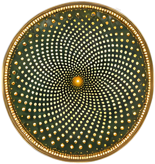
about 1,400 areolae at 137.508°
Diatoms wall themselves in two nested silica valves and do something like a fifth of Earth’s photosynthesis. This idealized valve places about 1,400 pores on Vogel’s sunflower spiral, each a further 137.508° around; the rim spirals count 55, 89 and 144.
KUNSTFORMEN
Caspar–Klug Capsid Geometry
12 five-fold vertices, always
Sixty subunits is the most an icosahedron can seat identically. Caspar and Klug’s 1962 fix: twelve pentamers at the vertices, hexamers between. Virologists quote triangulation numbers like shoe sizes; this is T = 4, like hepatitis B core.
KUNSTFORMEN
Bacteriophage T4
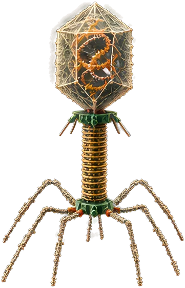
169 kb · 6 tail fibers
T4 looks like a lunar lander and lands like one. When its six long tail fibers bind, the baseplate flips from hexagon to star and the sheath contracts, driving the tube through the envelope to deliver 169 kb of DNA.
KUNSTFORMEN
Branching Colony Growth
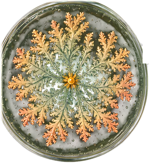
fractal dimension 1.72
Starve Bacillus subtilis on hard agar and the colony branches, and the branches branch. Nutrient diffuses in and the outermost tips catch most of it: diffusion-limited aggregation, fractal dimension about 1.7. The simulation behind this one measures 1.72.
THE PROGRAM
The Genetic Code
4³ = 64 codons
UUU was the first codon cracked, by Nirenberg and Matthaei in 1961. This wheel is computed from the standard table, so it is exact: 64 codons, three stops, AUG to start. Not quite universal: Mycoplasma reads UGA as tryptophan.
THE PROGRAM
The CRISPR Array
29 nt repeats · 32 nt spacers
Ishino found these 29-nucleotide repeats in E. coli in 1987 and could not say what they were for. They are immune memory: each 32-nucleotide spacer is a piece of a past invader, filed beside the leader, newest first.
THE PROGRAM
Cooper–Helmstetter Multifork Replication
ori / ter = 2^(C/τ) = 4
Copying the E. coli chromosome takes about 40 minutes, and division comes 20 minutes later, yet in rich medium cells divide every 20. Cooper and Helmstetter’s 1968 answer: start new rounds before old ones finish. Hence four origins, one terminus.
THE PROGRAM
The Genetic Toggle Switch
two stable states
Two repressors, each silencing the other’s gene. In 2000 Gardner, Cantor and Collins derived the condition for bistability from a two-variable model, then built plasmids to meet it. A pulse of inducer flips the switch; it stays flipped.
THE PROGRAM
Min Protein Oscillation
one cycle in 58 s
E. coli finds its middle with an oscillator, caught by Raskin and de Boer in 1999. MinD caps one pole until MinE strips it off, then re-forms opposite, about once a minute; MinC rides along, leaving mid-cell free to divide.
LOW REYNOLDS
The Flagellar Motor
11 stators · ~300 Hz
A proton-driven rotary motor: eleven stators turn a rotor that spins the flagellum at up to about 300 revolutions a second.
LOW REYNOLDS
Run-and-Tumble Chemotaxis
2.7 µm/s up the gradient
E. coli is too small to compare concentrations across its own body, so it compares over time: runs of about a second, tumbles averaging 68 degrees, fewer tumbles when things improve. Berg and Brown tracked it in 1972. The E. coli behind this page run and tumble 25 times slower than life.
LOW REYNOLDS
ATP Synthase Rotary Stepping
80° + 40° substeps
Boyer proposed that ATP synthase turns; in 1997 Noji and colleagues watched F1 spin an actin filament. Each 120-degree step uses one ATP: 80 degrees on binding, 40 more after a two-millisecond catalytic dwell, at close to full efficiency.
LOW REYNOLDS
Kirby–Bauer Disk Diffusion
24 mm zone at MIC 1 µg/mL
Standardized in 1966, run daily: a lawn on Mueller–Hinton agar 4 mm deep, antibiotic disks, a ruler. Zone edges freeze where diffusing drug meets the MIC; in the model, a 30 µg disk gives 24 mm at MIC 1 µg/mL.
LOW REYNOLDS
Michaelis–Menten and Lineweaver–Burk
v = Vmax[S] / (Km + [S])
Michaelis and Menten’s 1913 hyperbola: at Km half the enzyme is busy, and extra substrate buys ever less. The spheres count it, 2, 4, 7 of 8. Lineweaver and Burk straightened it in 1934; kineticists still argue about that plot.
MID-LOG
Monod Kinetics and the Growth Curve
lag · log · stationary · death
Monod called culture growth the basic method of microbiology; his growth-rate hyperbola looks like Michaelis–Menten but is empirical. The flasks sample one culture through lag, log, stationary and death. Diauxie, a two-step version, led him to the lac operon.
MID-LOG
The Bacterial Growth Laws
42% ribosomes at 20 min
Ribosomes make all protein, including ribosomes, so a faster-growing cell must devote more of its proteome to them. Scott, Hwa and colleagues made that a linear law in 2010. At a 20-minute doubling it puts about 42% there.
MID-LOG
The Quorum-Sensing Switch
on at 2.30, off at 1.81
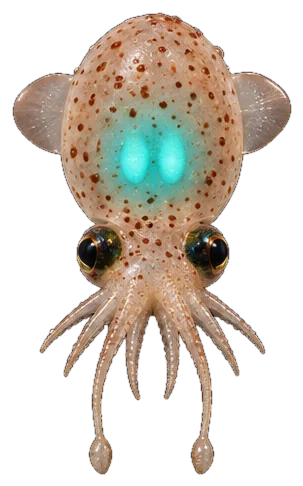
Vibrio fischeri glows in the bobtail squid’s light organ, never in open seawater. Its signal, an acyl-homoserine lactone, induces its own synthesis, so the population switches on at one density and off only at a lower one.
MID-LOG
Phage-Bacteria Predator-Prey Cycles
T₀ = 2π / √(rm)
Campbell wrote the equations in 1961: Lotka–Volterra, with adsorption rate, burst size and phage loss as the constants. Bacteria bloom, phage follow, bacteria crash, phage starve. In the ocean the same arithmetic kills roughly a fifth of bacterial biomass daily.
MID-LOG
Thermal Death Kinetics and the 12-D Cook
log₁₀ N/N₀ = −t / D
Heat kills spores first-order: each D-value at fixed temperature removes nine in ten. Esty and Meyer measured botulinum spores in 1922; the canner’s 12-D cook, 2.52 minutes at 121.1 °C, is usually rounded up to 3.
GRAM-VARIABLE
The Luria–Delbrück Fluctuation Test
σ² / ⟨r⟩ ≫ 1
Luria saw the test watching a colleague win at a slot machine. Induced resistance predicts Poisson counts; he and Delbrück found jackpots in 1943, variances far above the means. Mutation comes first, selection second.
GRAM-VARIABLE
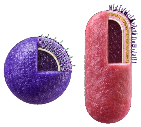
Gram-Positive and Gram-Negative Envelopes
thick wall vs outer membrane
Gram’s 1884 stain reads the envelope. Gram-positive: one membrane under tens of nanometers of peptidoglycan, which holds the dye. Gram-negative: a few nanometers of wall in the periplasm, beneath an outer membrane faced with lipopolysaccharide, whose lipid A is endotoxin.
GRAM-VARIABLE
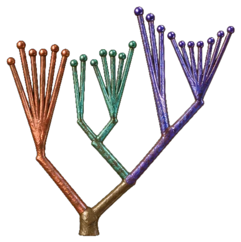
Woese’s Three Domains
6 + 7 + 10, as drawn in 1990
Ribosomal RNA split life into Bacteria, Archaea and Eukarya, with the archaea closer to us than to bacteria.
GRAM-VARIABLE
GC Skew and the Origin of Replication
s = (G − C) / (G + C)
The two strands are copied differently, and over millions of generations the leading strand gathers more G than C. The skew flips at origin and terminus, so its running sum draws a V whose minimum finds oriC in new genomes.
GRAM-VARIABLE
Poisson Infection and the MOI
63.2% infected at MOI 1
Viruses land on cells at random, so at an MOI of 1, 36.8% of cells get none, 36.8% one and 26.4% two or more. Pooled lentiviral screens therefore go in near 0.3, so most infected cells carry one construct.